Dosya:Tetraspora gelatinosa.jpg

Tam çözünürlük ((1.047 × 700 piksel, dosya boyutu: 139 KB, MIME tipi: image/jpeg))
Bu dosya Wikimedia Commons'ta bulunmaktadır. Dosyanın açıklaması aşağıda gösterilmiştir. Commons, serbest/özgür telifli medya dosyalarının bulundurulduğu depodur. Siz de yardım edebilirsiniz. |
Özet
| Açıklama |
Identifier: algvolimyxophy00west Title: Algæ. Vol. I. Myxophyceæ, Peridinieæ, Bacillarieæ, Chlorophyceæ, together with a brief summary of the occurrence and distribution of freshwat4er Algæ Year: 1916 (1910s) Authors: West, G. S. (George Stephen), 1876-1919 Subjects: Algae Publisher: Cambridge [Eng.] The University press Contributing Library: MBLWHOI Library Digitizing Sponsor: MBLWHOI Library
Click here to view book online to see this illustration in context in a browseable online version of this book.
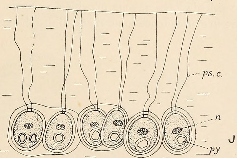
Text Appearing After Image: Fig. 113. A-G, Schizochlamys gelatinosa A. Br. A, vegetative cell showing pseudocilia, ×625; B, cell showing ecdysis of outer layers of wall, ×415; C and D, formation of zoogonidia, ×625; E, zoogonidium, ×830; F and G, zoogonidium changing to Schizochlamys-cell, ×830. H and I, Apiocystis brauniana Näg. H, pear-shaped colony, ×430; I, three cells showing pseudocilia, b, two daughter-cells from a division, the second pseudocilium not yet developed, ×860. J, Tetraspora gelatinosa (Vauch.) Desv., periphery of colony showing a few of the cells with their pseudocilia, × about 900. cv, contractile vacuole; n, nucleus; ol, oil globule; ps.c., pseudocilia; py, pyrenoid ; sf, stigma (or pigment-spot). (A-G, after Scherffel; J, after Chodat.) colony. The cells multiply by repeated division, chiefly in two directions inone plane, with the conversion of the walls of the mother-cells into mucilage.The pseudocilia are embedded in the mucilage of the colony (fig. 113 J), andeach cell is o
|
| Tarih | |
| Kaynak | Image from page 200 of "Algæ. Vol. I. Myxophyceæ, Peridinieæ, Bacillarieæ, Chlorophyceæ, together with a brief summary of the occurrence and distribution of freshwat4er Algæ" (1916) |
| Yazar | Internet Archive Book Images |
| İzin (Bu dosyanın tekrar kullanımı) |
Internet Archive Book Images @ Flickr Commons |
| Diğer sürümler |
.jpg)
Lisanslama
This image was taken from Flickr's The Commons. The uploading organization may have various reasons for determining that no known copyright restrictions exist, such as:
More information can be found at https://flickr.com/commons/usage/. Please add additional copyright tags to this image if more specific information about copyright status can be determined. See Commons:Licensing for more information. |
Lisanslama
|
Bu çalışma ABD'de veya yazarın yaşamının sona ermiş olmasından 100 veya daha fazla süre geçtiğinde bu duruma uygun telif yasaları olan tüm ülkelerde kamu malıdır. Bu çalışma 1 Ocak 1929 tarihinden önce yayımlanmıştır (veya ABD Telif Hakkı Ofisinde kayıtlıdır). Bu nedenle eser ABD'de kamu malıdır. | |
| Bu dosyanın, tüm ilgili ve komşu haklar da dâhil olmak üzere, telif hakkı yasası kapsamında bilinen kısıtlamalardan arınmış olduğu belirlendi. | |
Dosya geçmişi
Dosyanın herhangi bir zamandaki hâli için ilgili tarih/saat kısmına tıklayın.
| Tarih/Saat | Küçük resim | Boyutlar | Kullanıcı | Yorum | |
|---|---|---|---|---|---|
| güncel | 14.06, 3 Ekim 2019 | | 1.047 × 700 (139 KB) | Awkwafaba | File:Image from page 200 of "Algæ. Vol. I. Myxophyceæ, Peridinieæ, Bacillarieæ, Chlorophyceæ, together with a brief summary of the occurrence and distribution of freshwat4er Algæ" (1916).jpg cropped 35 % horizontally, 57 % vertically using CropTool with lossless mode. |
.jpg)
Dosya kullanımı
Bu görüntü dosyasına bağlantısı olan sayfalar:
Küresel dosya kullanımı
Aşağıdaki diğer vikiler bu dosyayı kullanır:
- ceb.wikipedia.org üzerinde kullanımı
- fr.wikipedia.org üzerinde kullanımı
- species.wikimedia.org üzerinde kullanımı
- www.wikidata.org üzerinde kullanımı
- zh.wikipedia.org üzerinde kullanımı