Dosya:FIPCytology2.jpg
FIPCytology2.jpg ((350 × 234 piksel, dosya boyutu: 40 KB, MIME tipi: image/jpeg))
Bu dosya Wikimedia Commons'ta bulunmaktadır. Dosyanın açıklaması aşağıda gösterilmiştir. Commons, serbest/özgür telifli medya dosyalarının bulundurulduğu depodur. Siz de yardım edebilirsiniz. |
Özet
| Açıklama |
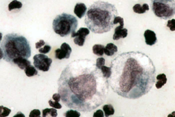
English: Color micrograph of the cytology of FIP-induced effusion. Magnification not specified; estimated to be 1000x.
Original caption: "The cytology of FIP effusion usually contains neutrophils, macrophages and lymphocytes." Image from "Feline Infectious Peritonitis: An Overview of Disease Transmission, Pathogenesis, Signs and Treatment With Emphasis on Diagnosis" ([1]) Clinical Pathology Clerkship Program |
| Tarih | 30 Eylül 2005 (original upload date) |
| Kaynak | en.wikipedia üzerinden Commons'a transfer edildi. |
| Yazar | The original uploader was Bk0 at İngilizce Vikipedi. |
Lisanslama

|
Bu dosyanın telif hakkı sahibi, telif hakkına uygun şekilde atıfta bulunmak koşuluyla herkese herhangi bir amaç için bu çalışmayı kullanmak üzere izin vermiştir. |
|
|
Orijinal yükleme günlüğü
- 2005-09-30 00:14 Bk0 350×234×8 (40687 bytes) Color micrograph of the cytology of [[Feline infectious peritonitis|FIP]]-induced effusion. Magnification not specified; estimated to be 1000x. Original caption: "The cytology of FIP effusion usually contains neutrophils, macrophages and lymphocytes." I
Dosya geçmişi
Dosyanın herhangi bir zamandaki hâli için ilgili tarih/saat kısmına tıklayın.
| Tarih/Saat | Küçük resim | Boyutlar | Kullanıcı | Yorum | |
|---|---|---|---|---|---|
| güncel | 18.53, 29 Aralık 2007 | | 350 × 234 (40 KB) | Euthygenes | {{Information |Description={{en|Color micrograph of the cytology of FIP-induced effusion. Magnification not specified; estimated to be 1000x. Original caption: "The cytology of FIP effusion usually contains neutrophi |
Dosya kullanımı
Bu görüntü dosyasına bağlantısı olan sayfalar:
Küresel dosya kullanımı
Aşağıdaki diğer vikiler bu dosyayı kullanır:
- el.wikipedia.org üzerinde kullanımı
- en.wikipedia.org üzerinde kullanımı
- et.wikipedia.org üzerinde kullanımı
- fr.wikipedia.org üzerinde kullanımı
- hu.wikipedia.org üzerinde kullanımı
- ko.wikipedia.org üzerinde kullanımı
- zh.wikipedia.org üzerinde kullanımı